

Have you ever wondered what kidney stones look like up close? These painful crystals can hurt our urinary system. Yet, under a microscope, they are diverse and complex. Let’s explore kidney stones and what they reveal about our health.

The Hidden Beauty of Kidney Stones
Kidney stones are painful but can be beautiful under magnification. They show off stunning crystals, bright colors, and unique patterns. They look more like art than medical samples.
Calcium oxalate kidney stone under microscope
Image: A 640px calcium oxalate kidney stone under magnification, showcasing its intricate crystalline structure.
Types of Kidney Stones and Their Microscopic Appearances
Kidney stones vary in type and appearance:
- Calcium Oxalate Stones: Common and jagged. They can be colorless, yellow, or light brown.
- Uric Acid Stones: Smooth and rounded, these range from yellow to reddish-brown.
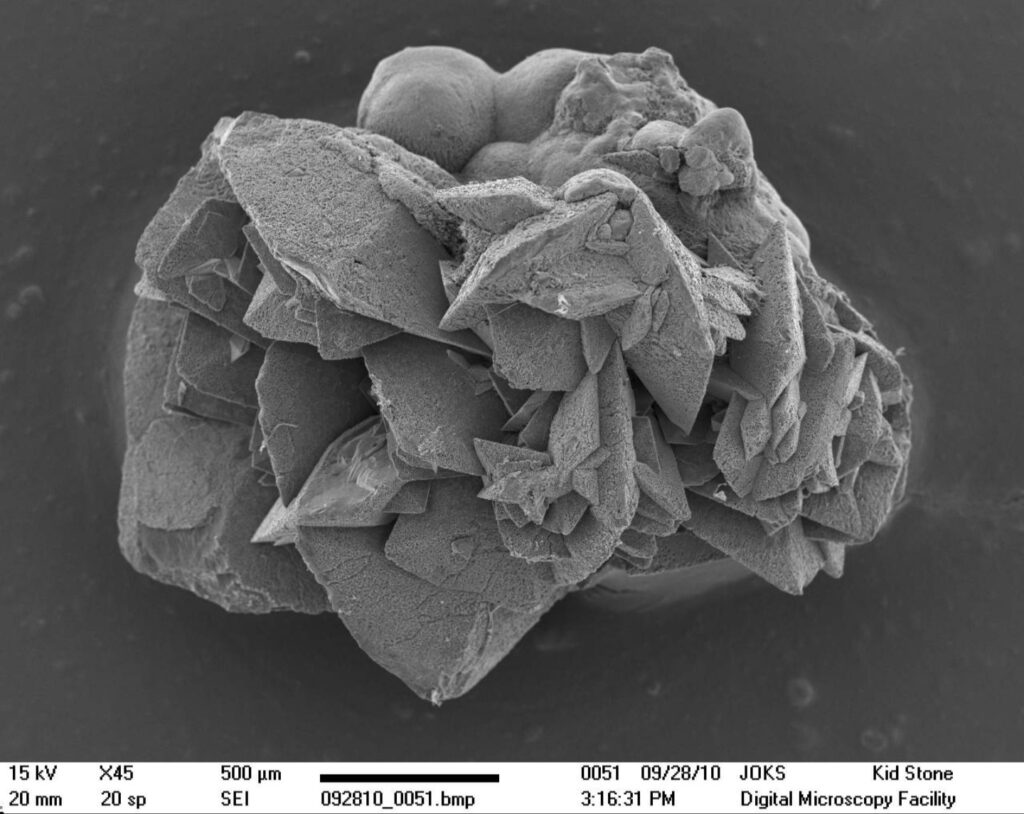
- Struvite Stones: Known as infection stones. They are larger, with a “coffin lid” shape, often white or tan.
Struvite kidney stone under microscope
Image: A struvite kidney stone showing its characteristic “coffin lid” shape under 200x magnification.
- Cystine Stones: The rarest type, these are flat, transparent, and hexagonal.
What Kidney Stones Reveal About Your Health
Looking at kidney stones under a microscope gives important health insights. Different features indicate various health issues:
- Crystal size and shape: Show how fast the stone formed and if it needs help to pass.
- Color variations: Suggest dietary or metabolic problems.
- Layering patterns: Reflect changes in urine composition.
- Presence of certain minerals: Point to metabolic disorders or nutritional imbalances.
The Importance of Stone Analysis
After passing a kidney stone, your doctor might ask you to collect it for analysis. This is crucial for several reasons:
- It helps identify the stone’s type, guiding treatment and prevention.
- It can reveal underlying health issues.
- It allows doctors to tailor dietary and medication advice.
Preventing Future Stones: Lessons from the Microscope
Studying kidney stones helps in prevention. For instance:
- Common calcium oxalate crystals suggest cutting back on certain foods.
- Uric acid stones indicate a need to lower purine intake.
- Struvite stones show a need for better urinary tract infection management.
In conclusion, examining kidney stones microscopically provides valuable prevention insights.han meets the eye!
FAQs About Kidney Stones Under the Microscope
1. Q: Can doctors determine the cause of kidney stones just by looking at them under a microscope?
A: While microscopic examination provides valuable clues, it’s usually combined with other tests and patient history to determine the exact cause of stone formation.
2. Q: Are kidney stones always visible under a regular microscope?
A: Most kidney stones can be seen under a standard light microscope, but some may require more advanced techniques like electron microscopy for detailed analysis.
3. Q: Can the microscopic appearance of a kidney stone predict how painful it will be to pass?
A: The size and shape of the stone, which can be observed microscopically, can give some indication of potential discomfort during passage. However, pain levels can vary greatly among individuals.