
Quick Summary
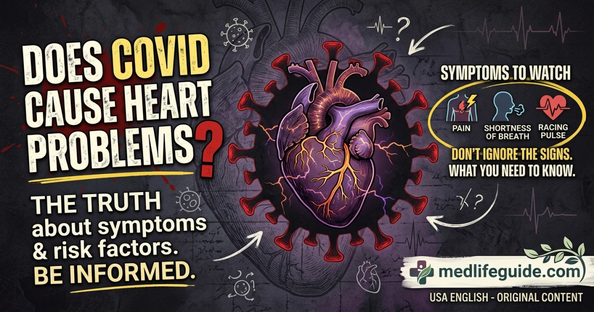
- Direct and Indirect Pathways: COVID-19 can cause cardiovascular issues through direct heart muscle inflammation (myocarditis), systemic blood clotting, and severe strain due to low blood oxygen levels.
- Broad Spectrum of Complications: Cardiovascular risks range from mild racing heart rate (tachycardia) and elevated blood pressure to severe conditions like heart failure, heart attacks, and arrhythmias.
- Long-Term Post-Acute Vulnerability: Heart complications can develop during the active viral infection or weeks to months later as part of Long COVID or Post-Acute Sequelae of SARS-CoV-2 (PASC).
EMERGENCY WARNING: If you or someone you are caring for experiences persistent chest pain or squeezing pressure, severe or sudden shortness of breath, sudden weakness or numbness on one side of the face or body, fainting or sudden loss of consciousness, or a pale, gray, or blue discoloration of the lips, face, or nail beds, call 911 (or your local emergency number) immediately. Do not wait to see if symptoms improve on their own.
1. Medical Overview and Pathophysiology
When SARS-CoV-2 enters the human body, it primarily targets respiratory tissue. However, clinical research has established that the virus can profoundly impact the cardiovascular system. Understanding how COVID-19 damages cardiac tissue involves examining several overlapping biological mechanisms, ranging from cellular invasion to widespread immune responses.
Cellular Entry and Direct Viral Invasion
The virus attaches to cells using its spike protein, which binds to angiotensin-converting enzyme 2 (ACE2) receptors. ACE2 receptors are present in pulmonary tissue and are also found in high concentrations on endothelial cells (the inner lining of blood vessels) and cardiomyocytes (heart muscle cells).
When SARS-CoV-2 directly infects these cardiac tissues, it can induce cellular injury, cellular death, and localized inflammation. Direct viral infection of cardiomyocytes can cause myocarditis, an inflammation of the heart muscle that impairs its ability to pump blood efficiently.
Cytokine Storm and Systemic Inflammation
In moderate to severe COVID-19 infections, the immune system may trigger an exaggerated inflammatory response known as a cytokine storm. High levels of pro-inflammatory proteins (such as Interleukin-6, Interleukin-1β, and Tumor Necrosis Factor-alpha) flood the bloodstream.
This systemic inflammation triggers widespread cellular stress throughout the cardiovascular system. It can weaken the heart muscle (inflammatory cardiomyopathy) and destabilize pre-existing fatty deposits (atherosclerotic plaques) in the coronary arteries, potentially leading to plaque rupture and acute myocardial infarction (heart attack).
Plaintext
SARS-CoV-2 Viral Infection
       │
       ├──► ACE2 Receptor Binding (Cardiomyocytes & Endothelium) ──► Direct Muscle Damage & Myocarditis
       │
       ├──► Systemic Immune Hyperactivation (Cytokine Storm)  ──► Vascular Inflammation & Plaque Rupture
       │
       └──► Endothelial Dysfunction & Hypercoagulability     ──► Microvascular Clots & Pulmonary Embolism
Endothelial Dysfunction and Blood Clotting (Hypercoagulability)
The endothelial cells lining the circulatory system help control blood vessel tone, clotting mechanisms, and vascular permeability. SARS-CoV-2 causes severe endothelial injury (endotheliitis), disrupting these natural protective functions.
Combined with systemic inflammation, this injury creates a hypercoagulable state—a condition where the blood becomes unusually prone to clotting. This can lead to small micro-clots in microscopic cardiac blood vessels, as well as larger blood clots such as deep vein thrombosis (DVT) and pulmonary embolism (PE). When pulmonary blood flow is restricted by a clot, the right ventricle of the heart must work significantly harder to pump blood through the lungs, leading to right-sided heart strain or failure.
Hypoxia, Demand Ischemia, and Autonomic Dysfunction
Severe lung involvement in COVID-19 frequently leads to low blood oxygen levels (hypoxia). Because the heart requires a continuous oxygen supply to function, low blood oxygen forces the heart muscle to beat faster and harder despite receiving reduced oxygen delivery. This imbalance between oxygen supply and demand leads to demand ischemia or Type 2 myocardial infarction.
Furthermore, COVID-19 can disrupt the autonomic nervous system, which regulates involuntary body processes like heart rate and blood pressure. This dysfunction, often called dysautonomia, can lead to persistent resting tachycardia (abnormally high heart rate) or Postural Orthostatic Tachycardia Syndrome (POTS), where the heart rate spikes dramatically upon standing.
2. Symptom Breakdown and Diagnostic Comparison
Distinguishing between primary respiratory COVID-19 symptoms, emerging cardiac complications, and non-COVID cardiovascular conditions is essential for early diagnosis and timely clinical care.
Common Cardiac Symptoms Related to COVID-19
- Palpitations: Sensations of a racing, pounding, fluttering, or skipping heart rate, often occurring at rest or with minimal exertion.
- Chest Tightness or Pain: A dull ache, sharp discomfort, or feeling of pressure in the middle or left side of the chest.
- Shortness of Breath (Dyspnea): Breathlessness that feels out of proportion to your activity level, or difficulty breathing while lying flat (orthopnea).
- Exertional Intolerance: Marked fatigue, dizziness, or weakness after light physical activity, such as walking up a single flight of stairs.
- Lightheadedness and Presyncope: Feeling faint or dizzy when standing up quickly due to blood pressure fluctuations or heart rhythm variations.
- Peripheral Edema: Swelling in the lower legs, ankles, or feet caused by fluid buildup, which can signal right-sided or congestive heart failure.
Symptom Differentiation Table
| Symptom / Feature | COVID-19 Cardiac Complication | Typical COVID-19 Respiratory | Non-COVID Heart Condition |
|---|---|---|---|
| Primary Chest Sensation | Squeezing, pressure, or sharp inflammation pain | Tightness secondary to coughing or deep breaths | Pressure, heaviness, or burning sensation |
| Shortness of Breath | Sudden worsening or persists when lying flat | Gradual, linked to nasal congestion or cough | Linked to physical effort or exertion |
| Heart Rate Pattern | Sudden spikes, fluttering, or persistent resting rate >100 bpm | Slightly elevated due to fever or dehydration | Irregular (e.g., AFib) or exertion-linked |
| Fever & Body Aches | Present during initial phase, may re-emerge later | Almost universally present during early infection | Typically absent unless infectious endocarditis |
| Leg Swelling (Edema) | Occurs if fluid accumulates from heart strain | Absent | Common in chronic heart failure or DVT |
| Response to Rest | Palpitations or pressure may persist at rest | Improves as body temperature cools and rest occurs | Angina often improves with rest or nitroglycerin |
3. Unique Clinical Takeaways
The following clinical insights reflect specialized observational data and emerging evidence regarding COVID-19 cardiovascular presentations:
- The “Biphasic” Cardiac Presentation in Post-Acute Recovery: Many patients recover from their initial mild or moderate respiratory illness, only to experience new-onset cardiovascular symptoms 2 to 6 weeks later. This delayed presentation is driven by persistent immune activation, autoimmune cross-reactivity, or lingering endothelial inflammation rather than active viral replication. Patients and caregivers should remain watchful for new chest discomfort, unexpected fatigue, or racing heart rate weeks after an initial COVID-19 infection has cleared.
- Postural Orthostatic Tachycardia Syndrome (POTS) and Dysautonomia Overlap: Up to 30% of individuals suffering from Long COVID report symptoms consistent with autonomic nervous system dysfunction, such as POTS. In these individuals, standing upright causes an abnormal heart rate increase (often exceeding 30 beats per minute above baseline) without a drop in blood pressure. This is frequently misdiagnosed as panic or anxiety, but it represents a structural or functional disturbance in autonomic vascular signaling following viral clearance.
- Subclinical Myocarditis in Young and Athletic Populations: Young, healthy adults and competitive athletes can develop mild, subclinical myocarditis (heart muscle inflammation with minimal or no overt symptoms during initial infection). Resuming vigorous, high-intensity aerobic exercise while the heart muscle is actively inflamed increases the risk of triggering severe ventricular arrhythmias. Clinical guidelines advise gradual, phased return-to-play protocols for athletes recovering from COVID-19.
4. Stage-by-Stage Illness Progression Timeline
Cardiovascular complications can manifest at different stages of a COVID-19 infection. The timeline below illustrates how heart involvement can develop over time.
Plaintext
[Days 1–7: Acute Phase] ────► [Days 8–14: Inflammatory Peak] ────► [Weeks 3–12+: Post-Acute Phase]
 Viral replication & fever       Cytokine surge, oxygen drops,      Immune dysregulation, POTS,
 High heart rate from fever      micro-clots & heart strain         persistent exercise intolerance
Stage 1: Acute Viral Phase (Days 1 to 7)
- Pathophysiological Activity: Active viral replication occurs in the upper and lower respiratory tracts. Mild systemic inflammation begins.
- Cardiovascular Symptoms: Sinus tachycardia (elevated heart rate) caused by fever, dehydration, and stress is common. Mild blood pressure fluctuations may occur.
- Primary Risks: In patients with advanced underlying heart disease, increased physical stress and fever can trigger early angina or demand ischemia.
Stage 2: Hyper-Inflammatory & Thrombotic Peak (Days 8 to 14)
- Pathophysiological Activity: Viral loads begin to drop, but the host immune response may become hyperactive. Cytokine levels peak, and risk of endothelial damage and hypercoagulability increases.
- Cardiovascular Symptoms: Onset of severe shortness of breath, sharp chest pain indicative of pericarditis or myocarditis, central chest pressure, or leg swelling.
- Primary Risks: Acute myocarditis, stress cardiomyopathy (Takotsubo syndrome), deep vein thrombosis, pulmonary embolism, or acute coronary syndrome.
Stage 3: Post-Acute Sequelae and Recovery Phase (Weeks 3 to 12+)
- Pathophysiological Activity: Active viral infection has resolved, but residual low-grade tissue inflammation, microvascular dysfunction, or autonomic disturbance may persist.
- Cardiovascular Symptoms: Lingering exercise intolerance, postural tachycardia (POTS), sudden heart rate spikes with minimal movement, persistent brain fog accompanied by lightheadedness, or ongoing chest tightness.
- Primary Risks: Chronic post-viral fatigue, persistent heart failure, long-term autonomic dysfunction, or delayed pericardial inflammation.
5. High-Risk Vulnerabilities and Special Populations
While COVID-19-related heart issues can affect people of any age, specific groups face distinct risks and challenges.
Plaintext
                 ┌─────────────────────────────────────────┐
                 │       High-Risk Patient Profiles        │
                 └────────────────────┬────────────────────┘
                                      │
     ┌────────────────────────────────┼────────────────────────────────┐
     ▼                                ▼                                ▼
Older Adults (65+)             Pediatric Patients              Immunocompromised &
• Pre-existing CAD/HF          • MIS-C (Multi-System           Pre-existing Cardiac
• Subtle confusion/lethargy      Inflammatory Syndrome)        • Reduced reserve capacity
• Atypical heart attacks       • Coronary artery aneurysms     • Delayed viral clearing
Older Adults (Aged 65 and Older)
Older individuals are more likely to have underlying cardiovascular conditions, such as coronary artery disease, hypertension, or heart failure. When exposed to COVID-19, their cardiovascular reserve capacity is lower, making them more susceptible to demand ischemia, acute heart failure exacerbation, and arrhythmias.
Furthermore, elderly patients may present with atypical symptoms; rather than complaining of sharp chest pain, they may show sudden confusion, acute lethargy, loss of appetite, or unexplained falls caused by temporary drops in blood pressure.
Pediatric Populations and MIS-C
Although children generally experience milder acute COVID-19 infections compared to adults, a small percentage can develop a serious post-infectious condition called Multisystem Inflammatory Syndrome in Children (MIS-C), typically occurring 2 to 6 weeks after infection.
MIS-C causes widespread inflammation in the heart, blood vessels, digestive tract, and other organs. Cardiac manifestations of MIS-C include severe left ventricular dysfunction, acute heart failure, myocarditis, and dilatation or aneurysms of the coronary arteries (similar to Kawasaki disease). Parents should watch for persistent high fever, bloodshot eyes, red or swollen palms and soles, abdominal pain, and extreme lethargy.
Individuals with Pre-existing Cardiovascular Disease
Patients with pre-existing heart failure, prior heart attacks, structural heart defects, or poorly controlled high blood pressure face higher rates of hospitalization and complications if infected with COVID-19. The added metabolic and oxygen demand caused by viral infection can convert stable heart disease into decompensated heart failure. Strict compliance with prescribed cardiac medications during viral illness is vital for these patients.
6. Diagnostic Evaluation and Medical Management Guidelines
If a healthcare provider suspects COVID-19-related heart involvement, they will conduct a structured diagnostic evaluation and implement evidence-based therapies.
Diagnostic Testing
- High-Sensitivity Cardiac Troponin (hs-cTn): A blood test measuring troponin, a protein released when heart muscle cells are damaged. Elevated levels indicate myocardial injury from myocarditis, heart attack, or severe strain.
- B-Type Natriuretic Peptide (BNP or NT-proBNP): A biomarker released by the heart in response to increased wall pressure and fluid overload. Elevated levels suggest worsening heart failure or significant cardiac strain.
- Electrocardiogram (ECG / EKG): Records the electrical activity of the heart to detect arrhythmias, heart blockages, signs of acute ischemia, or pericarditis (evidenced by widespread ST-segment elevation or PR depression).
- Echocardiogram: An ultrasound of the heart that visualizes heart structure, valve function, wall movement, and overall pumping efficiency (ejection fraction).
- Cardiac Magnetic Resonance Imaging (Cardiac MRI): The gold standard non-invasive imaging test for diagnosing myocarditis. It can detect tissue swelling (edema), inflammation, and scar tissue (late gadolinium enhancement) within the heart muscle.
- Inflammatory and Coagulation Markers: C-reactive protein (CRP), Erythrocyte Sedimentation Rate (ESR), and D-dimer blood tests help assess systemic inflammation and blood clotting activity.
Medical Management Protocols
- Antiviral Therapies: Administering approved oral or intravenous antivirals (such as Paxlovid or Remdesivir) early in the acute infection phase reduces viral replication, decreasing systemic strain and downstream cardiac complications in eligible high-risk individuals.
- Anticoagulation Therapy: Hospitalized patients with elevated clotting markers or severe COVID-19 are frequently given preventive (prophylactic) or therapeutic doses of blood thinners (heparin) to lower the risk of blood clots and micro-thrombosis.
- Anti-Inflammatory Medications: In patients with severe systemic inflammation or myocarditis, physicians may prescribe corticosteroids (such as dexamethasone) or targeted immunomodulatory agents to reduce cardiac tissue injury.
- Heart Failure and Arrhythmia Treatments: Standard guideline-directed medical therapy for heart failure—including ACE inhibitors/ARBs, beta-blockers, aldosterone antagonists, and diuretics—is initiated or continued as tolerated to support cardiac function.
7. Home Care, Protection Protocols, and Recovery
Managing recovery safely requires balancing rest, physical activity, and symptom monitoring.
Plaintext
┌────────────────────────────────────────────────────────────────────────┐
│                   Home Recovery Protocol Essentials                     │
├───────────────────────────────────┬────────────────────────────────────┤
│ • Pulse Oximeter Monitoring       │ Check SpO2 regularly (Target ≥95%) │
│ • Pacing & Activity Grading       │ Avoid sudden physical exertion     │
│ • Hydration & Electrolytes        │ Supports blood volume & pressure   │
│ • Follow-up Medical Visits        │ Re-evaluate persistent symptoms    │
└───────────────────────────────────┴────────────────────────────────────┘
Home Monitoring Tools and Guidance
- Pulse Oximetry: Monitor blood oxygen saturation (SpO2) at rest. Readings consistently below 95% should be promptly reported to a physician.
- Blood Pressure and Heart Rate Logs: Keep a daily record of morning and evening resting heart rate and blood pressure to share with your medical team.
- Hydration Management: Maintain adequate fluid intake (unless advised to restrict fluids by a physician due to heart failure) to preserve blood volume and lower the risk of postural lightheadedness.
Safe Return to Exercise (Pacing)
Returning to physical activity after COVID-19 infection should be gradual, especially if you experienced cardiac or respiratory symptoms.
- Avoid Early Vigorous Exercise: Refrain from high-intensity cardio, heavy lifting, or competitive sports during the acute infection phase and for at least 7 to 10 days after all symptoms resolve.
- Follow a “Pacing” Strategy: Slowly increase daily light walking while keeping your heart rate within a comfortable range. If you experience chest discomfort, palpitations, extreme fatigue, or shortness of breath, stop exercising immediately and rest.
- Medical Clearance: Individuals with pre-existing heart conditions, severe COVID-19 illness, or persistent cardiac symptoms should receive clearance from a physician or cardiologist before resuming strenuous exercise programs.
Infection Prevention and Community Protection
Protecting yourself and others from repeat infections helps prevent additive cardiovascular strain.
- Vaccination: Stay up to date with recommended COVID-19 vaccines, which significantly reduce the risk of severe disease, hospitalization, myocarditis, and long-term cardiac complications.
- Ventilation and Masking: Improve indoor air circulation and consider wearing a well-fitting, high-filtration mask (such as an N95 or KN95) in crowded or poorly ventilated public spaces during local respiratory virus surges.
- Hand Hygiene: Wash hands regularly with soap and water or use an alcohol-based hand sanitizer to minimize viral exposure.
Frequently Asked Questions (FAQs)
Yes, mild COVID-19 infections can occasionally lead to cardiac symptoms like palpitations, mild myocarditis, or autonomic dysregulation (POTS) during recovery.
It is impossible to reliably differentiate chest pain sources without diagnostic medical testing; any persistent chest pain or pressure requires immediate emergency medical evaluation.
Sinus tachycardia (an unusually fast resting heart rate) and temporary arrhythmias are the most frequently reported cardiac signs, followed by myocarditis and heart strain.
For many individuals, post-COVID palpitations gradually improve over several weeks to months with proper rest, hydration, and medical guidance.
Yes, vaccination lowers the risk of severe COVID-19 infection, which directly reduces the likelihood of developing severe cardiac issues, micro-clots, and Long COVID complications.
About the Reviewer
Dr. Adam N. Khan, MD is a board-certified internal medicine physician dedicated to patient education and evidence-based clinical guidance. He maintains no commercial conflicts of interest or financial disclosures related to cardiovascular therapies, medical devices, or pharmaceutical products.