
Medically Reviewed and Compiled by: Dr. Adam N. Khan, MD
Quick Summary
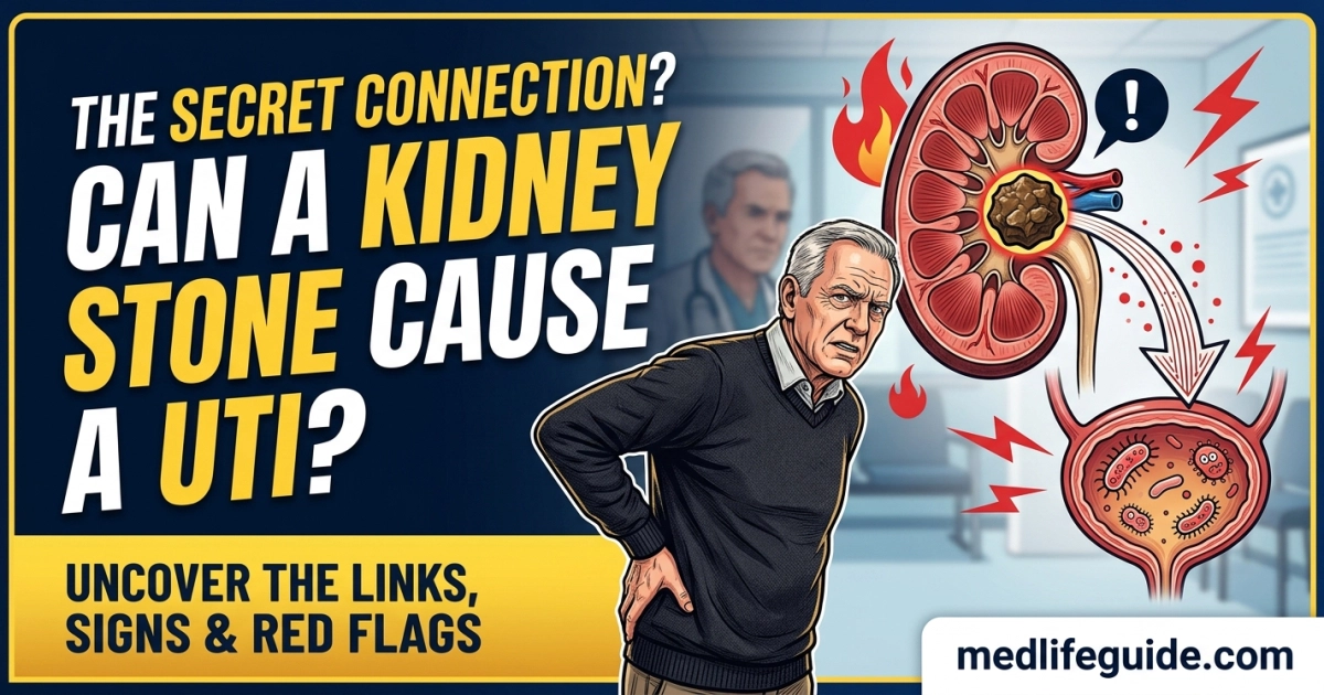
- Direct Cause: Kidney stones can directly cause urinary tract infections (UTIs) by blocking urine flow, causing fluid stagnation, damaging tissue linings, and harboring persistent bacterial biofilms.
- Two-Way Relationship: While stones can trigger infections, certain bacteria can also cause stones (struvite/infection stones), creating a dangerous cycle of chronic UTIs and rapid stone growth.
- Emergency Medical Red Flag: An obstructed kidney coupled with an active infection is a life-threatening medical emergency requiring urgent decompression to prevent urosepsis.
EMERGENCY WARNING
If you experience severe side or back pain accompanied by a high fever, uncontrollable shaking or chills, nausea and vomiting, confusion, or an inability to pass urine, call 911 or go to the nearest emergency room immediately. A kidney stone blocking infected urine is a severe emergency that can lead to rapid organ failure and urosepsis without immediate care.
1. Medical Overview and Pathophysiology
The relationship between kidney stones and urinary tract infections (UTIs) is complex and closely connected. To understand how a hard mineral deposit can lead to a bacterial infection, it helps to look at the normal function of the human urinary system.
The urinary tract consists of two kidneys, two ureters (thin tubes connecting the kidneys to the bladder), a bladder, and a urethra. Under healthy conditions, the continuous, downward flow of urine acts as a natural flushing mechanism. It prevents bacteria from traveling up the system and establishing colonies. However, when a mineral mass forms inside the kidney and migrates down into the narrow passages of the ureter or bladder, this protective system breaks down.
+-------------------------------------------------------------------------+
|                        MECHANISMS OF INFECTION                          |
+-------------------------------------------------------------------------+
|                                                                         |
|  1. URINARY OBSTRUCTION                                                 |
|     Stone lodges in ureter/bladder -> Urine flow stops -> Fluid pools  |
|                                                                         |
|  2. BACTERIAL MULTIPLICATION                                            |
|     Stagnant urine creates an ideal breeding ground for pathogens       |
|                                                                         |
|  3. TISSUE TRAUMA                                                       |
|     Jagged stone edges scratch mucous lining -> Creates entry points    |
|                                                                         |
|  4. BIOFILM FORMATION                                                   |
|     Bacteria coat the stone's surface -> Protected from antibiotics    |
|                                                                         |
+-------------------------------------------------------------------------+
Fluid Stagnation and Bacterial Growth
When a kidney stone partially or completely blocks the ureter or bladder outlet, urine cannot drain properly. This condition, known as urinary stasis, causes urine to back up into the renal pelvis and kidney tissue. Stagnant urine acts as a nutrient-rich culture medium. Bacteria such as Escherichia coli, Proteus mirabilis, or Klebsiella pneumoniae—which are normally flushed out during urination—can multiply rapidly in stagnant pools of urine, turning a mild bacterial presence into a full-blown UTI.
Epithelial Micro-Trauma and Mucosa Damage
Kidney stones are rarely smooth; they often possess sharp, crystalline structures. As a stone shifts, travels, or becomes wedged within the urinary tract, its abrasive surface scratches and tears the delicate mucosal lining (the urothelium). This micro-trauma triggers local inflammation, swelling, and microscopic bleeding. The damaged tissue loses its natural protective barriers, creating easy entry points for bacteria to attach, penetrate deeper into tissue layers, and cause infection.
Bacterial Biofilms and Foreign Body Effect
In the human body, a kidney stone acts as a non-living foreign object. Bacteria can adhere to the rough outer surface and internal microscopic pores of the stone. Once attached, these microorganisms produce a sticky protective matrix composed of proteins and sugars, known as a biofilm. Biofilms shield the underlying bacteria from both the body’s immune cells and circulating oral antibiotics. As long as the stone remains inside the body, it can act as a persistent reservoir for bacteria, frequently shedding live organisms into the urine and causing recurrent UTIs that return shortly after an antibiotic course is completed.
The Two-Way Cycle: Infection Stones (Struvite)
While kidney stones can cause UTIs, specific UTIs can also create kidney stones. This reciprocal relationship is most evident in the formation of struvite stones, also known as “infection stones.”
+-------------------------------------------------------------------------+
|                     STRUVITE STONE FORMATION CYCLE                      |
+-------------------------------------------------------------------------+
|                                                                         |
|  Urease-Producing Bacteria (e.g., Proteus) Infection                    |
|                         │                                               |
|                         ▼                                               |
|  Splits Urea into Ammonia (NH3) & Carbon Dioxide                        |
|                         │                                               |
|                         ▼                                               |
|  Urine pH Rises (Becomes Strongly Alkaline: > 7.2)                     |
|                         │                                               |
|                         ▼                                               |
|  Precipitation of Magnesium, Ammonium, and Phosphate                    |
|                         │                                               |
|                         ▼                                               |
|  Rapid Growth of Large Struvite / Staghorn Calculi                     |
|                         │                                               |
|                         └───────────────► (Cycle Repeats)               |
+-------------------------------------------------------------------------+
Struvite stones form exclusively when the urinary tract is infected with urease-producing bacteria, most commonly Proteus mirabilis, Klebsiella, or Pseudomonas. These specific bacteria produce an enzyme called urease, which hydrolyzes urea in the urine into ammonia and carbon dioxide. Ammonia elevates urine pH, transforming normally acidic urine into an alkaline environment (often with a pH greater than 7.2).
Under these alkaline conditions, magnesium, ammonium, and phosphate ions precipitate out of solution, forming magnesium ammonium phosphate hexahydrate crystals. These crystals aggregate rapidly around the bacterial colonies, forming large, branched structures known as “staghorn calculi” that can fill the entire renal collecting system.
2. Symptom Breakdown and Diagnostic Comparison
Because kidney stones and urinary tract infections affect the same organ system, their symptoms frequently overlap. Patients often experience a combination of severe localized discomfort and lower urinary tract symptoms. Distinguishing between an uncomplicated kidney stone, an uncomplicated lower UTI, and a combined obstructive infection is essential for proper medical care.
+-------------------------------------------------------------------------+
|                     SYMPTOM LOCATION CHARACTERISTICS                    |
+-------------------------------------------------------------------------+
|                                                                         |
|  KIDNEY STONE PAIN (Renal Colic)                                        |
|  - Sharp, intense, spasmodic pain in the flank/back                     |
|  - Radiates downward toward lower abdomen, groin, and testicles/labia   |
|  - Patients cannot sit still (the "kidney stone dance")                 |
|                                                                         |
|  LOWER UTI (Cystitis)                                                   |
|  - Dull, aching pain or pressure in the suprapubic area (lower belly)   |
|  - Burning sensation during urination (dysuria)                         |
|  - Constant, urgent need to empty bladder                               |
|                                                                         |
|  UPPER UTI / COMBINED OBSTRUCTION (Pyelonephritis / Infected Stone)     |
|  - Severe flank pain combined with systemic signs (fever, chills)       |
|  - Nausea, vomiting, dark/cloudy/foul-smelling urine                    |
|  - Requires immediate medical evaluation                                |
|                                                                         |
+-------------------------------------------------------------------------+
Primary Symptom Overlap
- Dysuria (Painful Urination): Both conditions irritate the urethral and bladder walls, causing a sharp burning sensation during urination.
- Urinary Frequency and Urgency: A stone lodged in the lower ureter near the bladder wall causes physical irritation that tricks the bladder into feeling full, mimicking the urgent, frequent urge to urinate caused by bacterial cystitis.
- Hematuria (Blood in Urine): Stones scratch the urinary tract lining, causing blood in the urine that may appear pink, red, or cola-colored. UTIs also cause inflammation that can lead to visible or microscopic bleeding.
- Cloudy or Odorous Urine: Pyuria (pus and white blood cells in the urine) occurs in active UTIs and can also happen when a stone causes significant local inflammation.
Diagnostic Comparison Table
| Clinical Feature | Uncomplicated Kidney Stone | Uncomplicated Lower UTI (Cystitis) | Combined Condition (Infected Obstructing Stone) |
| Primary Pain Site | Severe flank/back pain radiating to groin | Suprapubic (lower belly) discomfort/pressure | Severe flank pain WITH abdominal/suprapubic tenderness |
| Pain Pattern | Sharp, spasmodic waves (renal colic); patient writhes | Constant burning during urination and dull lower ache | Intense, relentless back/flank pain with systemic body aches |
| Fever & Chills | Unlikely (unless infected) | Absent or low-grade (below 100.4°F) | High fever (>101°F), shaking chills, night sweats |
| Nausea & Vomiting | Common (due to shared nerve pathways) | Rare | Very Common (persistent and severe) |
| Urine Urgency/Frequency | Present if stone is near bladder | Universal hallmark symptom | Very High, often with inability to pass urine |
| Urinalysis Findings | Microscopic/gross blood; normal white cells; negative nitrites | High white blood cells, positive nitrites, positive leukocyte esterase | Abundant white blood cells, bacteria, positive nitrites, micro/gross blood |
| Blood Tests (CBC) | White blood cell count usually normal | WBC count normal or mildly elevated | Marked leukocytosis (elevated WBC count) with left shift |
| Urgency Level | Urgent (Pain management and outpatient urology) | Moderate (Outpatient antibiotics within 24-48 hrs) | CRITICAL EMERGENCY (Immediate ER visit required) |
3. Unique Clinical Takeaways
To help patients and caregivers navigate this complex health issue, three critical clinical insights go beyond basic symptom lists:
- Takeaway 1: The “Silent” Obstructive Infection Window: In many medical conditions, pain serves as a reliable alarm system. However, when a kidney stone completely blocks a ureter, urine production in that specific kidney can drop due to high back-pressure within hours. If the kidney stops producing urine on the obstructed side, the sharp spasmodic pain (renal colic) may temporarily lessen or vanish, giving a false sense of recovery. Meanwhile, bacteria trapped behind the blockage continue to multiply rapidly in the trapped fluid. Patients may mistake this sudden drop in pain as a sign that the stone has passed, delaying care until they develop high fever, confusion, and systemic sepsis. A sudden disappearance of renal pain without a confirmed stone passing in the toilet—especially when accompanied by fatigue or low-grade fever—requires immediate medical evaluation.
- Takeaway 2: False Negatives in Standard Urine Cultures: Standard urine cultures rely on a sample collected from the bladder (a mid-stream voided specimen). If a kidney stone completely blocks one ureter, the urine reaching the bladder comes entirely from the healthy, unblocked kidney. As a result, a standard urine culture may return completely negative for bacteria, even while a severe, high-pressure infection is growing behind the stone in the opposite kidney. Clinicians who suspect an infected obstructing stone must look beyond simple urine dipsticks and rely on advanced imaging (such as non-contrast CT) and blood cultures to detect hidden infections.
- Takeaway 3: Post-Lithotripsy Bacterial Release: When medical procedures like Extracorporeal Shock Wave Lithotripsy (ESWL) or laser ureteroscopy break up a large kidney stone—particularly a struvite or chronic calcium phosphate stone—they shatter the outer shell of the stone. If bacteria are trapped deep within the layers of the stone, pulverizing it can suddenly release millions of viable bacteria and endotoxins directly into the urinary tract and bloodstream. This sudden release can trigger a rapid inflammatory response, causing fever, severe shivering, or septic shock within 12 to 24 hours after the procedure. Patients undergoing stone-breaking procedures should be carefully monitored for post-procedure infections and may require pre-procedure antibiotic treatment.
4. Illness Progression Timeline
Understanding how a kidney stone and a UTI interact over time helps patients and caregivers recognize when a routine issue is turning into a serious complication.
+-------------------------------------------------------------------------+
|                       STAGES OF DISEASE PROGRESSION                     |
+-------------------------------------------------------------------------+
|                                                                         |
|  STAGE 1: Stone Formation & Early Movement (Days 1–3)                   |
|  - Crystallization in kidney; silent movement down the ureter           |
|  - Onset of sharp, intermittent flank pain and mild blood in urine      |
|                                                                         |
|  STAGE 2: Obstruction & Urinary Stagnation (Days 3–7)                   |
|  - Stone lodges in narrow ureter junction; urine backs up               |
|  - Stagnant fluid allows bacteria to colonize and grow                  |
|                                                                         |
|  STAGE 3: Acute Combined Infection (Days 7–10)                          |
|  - Infection spreads to kidney tissue (pyelonephritis)                  |
|  - High fever, shaking chills, severe nausea, and cloudy urine appear    |
|                                                                         |
|  STAGE 4: Critical Complications or Resolution (Day 10+)                |
|  - Without care: Risk of urosepsis, perinephric abscess, kidney damage   |
|  - With care: Emergency drainage, antibiotic therapy, stone removal     |
|                                                                         |
+-------------------------------------------------------------------------+
Stage 1: Stone Formation and Early Movement (Days 1 to 3)
- Physiological Events: Mineral crystals precipitate out of supersaturated urine inside the renal calyces, forming a solid mass. The stone eventually dislodges and enters the narrow ureter.
- Clinical Symptoms: Sharp, sudden, localized pain in the back or side below the ribs. Pain comes in waves as the ureter contracts to push the stone forward. Urine may appear light pink or tea-colored due to minor scratches in the lining.
- Intervention Goal: Hydration, pain management with nonsteroidal anti-inflammatory drugs (NSAIDs) or alpha-blockers under medical supervision, and catching passed stones in a strainer.
Stage 2: Obstruction and Urinary Stagnation (Days 3 to 7)
- Physiological Events: The stone becomes lodged at a natural narrowing point, such as the ureteropelvic junction or ureterovesical junction. Urine cannot pass, causing the ureter and renal pelvis to swell (hydronephrosis). Bacteria entering from the lower tract colonize the stagnant pool.
- Clinical Symptoms: Constant, dull ache in the flank punctuated by sharp spasms. Bladder symptoms like frequency, burning, and urgency increase. Urine may turn cloudy or develop a strong, unpleasant odor.
- Intervention Goal: Medical evaluation including a non-contrast CT scan to confirm stone location and size. Urinalysis to check for early signs of infection.
Stage 3: Acute Combined Infection (Days 7 to 10)
- Physiological Events: Bacteria multiply rapidly in the obstructed kidney, leading to tissue invasion, micro-abscess formation, and acute pyelonephritis. High pressure pushes bacteria and their toxins across thin membrane barriers directly into the bloodstream.
- Clinical Symptoms: Sudden onset of high fever (above 101°F), violent shaking chills (rigors), severe nausea, vomiting, and profound fatigue. Pain becomes severe and constant across the back and abdomen.
- Intervention Goal: Emergency medical evaluation. Immediate hospital admission for intravenous (IV) antibiotics and urgent surgical decompression.
Stage 4: Critical Complications or Resolution (Day 10+)
- Physiological Events: Without rapid medical treatment, pressure and infection can destroy nephrons (functional kidney units), cause a collection of pus around the kidney (perinephric abscess), or trigger widespread systemic inflammation (urosepsis). With timely treatment, the blockage is relieved, infection clears, and kidney function recovers.
- Clinical Symptoms: In severe cases: rapid heart rate, low blood pressure, confusion, and reduced urine output. In successfully treated cases: steady reduction in pain, fever resolution, and return of clear urine.
- Intervention Goal: Long-term urological follow-up, 24-hour urine metabolic evaluation, dietary adjustments, and preventive care.
5. High-Risk Vulnerabilities and Special Populations
Certain patient groups face significantly higher risks when dealing with kidney stones and urinary tract infections. In these individuals, symptoms can present atypically, and complications can progress rapidly.
+-------------------------------------------------------------------------+
|                      SPECIAL POPULATION RISK PROFILES                   |
+-------------------------------------------------------------------------+
|                                                                         |
|  ELDERLY ADULTS (>65 YEARS)                                             |
|  - Masked symptoms: Low or absent fever due to blunted immune response  |
|  - Primary signs: Sudden confusion (delirium), lethargy, falls          |
|  - High risk of rapid progression to septic shock                       |
|                                                                         |
|  PREGNANT WOMEN                                                         |
|  - Increased risk: Progesterone relaxes ureters; physical compression   |
|  - Diagnostic challenge: Radiation limits require ultrasound imaging    |
|  - Risks: Preterm labor, low birth weight, acute pyelonephritis         |
|                                                                         |
|  PEDIATRIC PATIENTS                                                     |
|  - Hard-to-spot signs: Irritability, poor feeding, unexplained fever    |
|  - Under-recognized: Often linked to underlying structural anomalies    |
|                                                                         |
|  IMMUNOCOMPROMISED & DIABETIC PATIENTS                                  |
|  - High risk: Fungal infections, emphysematous pyelonephritis           |
|  - Impaired neutrophils allow rapid bacterial spread                    |
|                                                                         |
+-------------------------------------------------------------------------+
Older Adults (Aged 65 and Above)
Elderly individuals often have altered immune responses, making traditional signs of infection less obvious. They may not run a high fever, even during a severe kidney infection. Instead of classic flank pain, an older adult with an infected kidney stone might present with:
- Sudden onset of confusion, disorientation, or acute delirium
- Unexplained fatigue, lethargy, or weakness leading to falls
- Loss of appetite and reduced oral fluid intake
- Incontinence or sudden worsening of baseline urinary habits
Because these symptoms are easily attributed to dementia, aging, or general frailty, diagnosis is often delayed, placing older adults at a higher risk for urosepsis.
Pregnant Individuals
Pregnancy brings physiological changes that increase the risk of both kidney stones and UTIs. High levels of progesterone relax the smooth muscle walls of the ureters, slowing urine flow. At the same time, the expanding uterus physically compresses the ureters, worsening urine stagnation. If a pregnant woman develops a kidney stone that blocks urine flow and causes an infection, it poses serious risks to both mother and fetus, including:
- Increased risk of acute pyelonephritis and maternal sepsis
- High rates of premature uterine contractions and preterm labor
- Challenges in diagnostic imaging, as standard CT scans are avoided to minimize fetal radiation exposure (renal ultrasonography is used instead)
Pediatric Patients
Although less common, kidney stones in children are increasing in frequency due to dietary factors, metabolic conditions, and structural anomalies of the urinary tract. Infants and young children cannot easily describe localized flank pain or burning during urination. Parents and caregivers should watch for:
- Unexplained, persistent fever without obvious respiratory symptoms
- Irritability, inconsolable crying, or abdominal guarding
- Poor feeding, vomiting, and failure to gain weight
- Foul-smelling, cloudy, or blood-stained diapers
Immunocompromised and Diabetic Individuals
Patients living with diabetes mellitus, chronic kidney disease, or conditions requiring immunosuppressive therapy (such as organ transplants or autoimmune treatments) face elevated risks. High blood sugar levels in diabetic patients impair white blood cell function, reducing the body’s ability to fight off bacterial growth in stagnant urine. Furthermore, diabetic patients with infected kidney stones are at higher risk for rare, life-threatening complications such as emphysematous pyelonephritis—a severe, gas-forming infection of the kidney tissue that requires emergency intervention.
6. Evidence-Based Diagnostics, Testing, and Management
Proper medical management requires identifying the presence, size, and location of the stone while evaluating for an active bacterial infection.
+-------------------------------------------------------------------------+
|                      DIAGNOSTIC & MANAGEMENT WORKUP                     |
+-------------------------------------------------------------------------+
|                                                                         |
|  1. LABORATORY DIAGNOSTICS                                              |
|     - Urinalysis: Check leukocyte esterase, nitrites, protein, blood    |
|     - Urine Culture & Sensitivity: Identify bacteria & choose drugs     |
|     - Serum CBC & Metabolic Panel: Evaluate WBC count & kidney function |
|     - Blood Cultures: Performed if high fever or sepsis is suspected    |
|                                                                         |
|  2. DIAGNOSTIC IMAGING                                                  |
|     - Non-Contrast CT (Abdomen/Pelvis): Gold standard for stone detection|
|     - Renal Ultrasound: First-line for pregnant women and children      |
|                                                                         |
|  3. URGENT SURGICAL DECOMPRESSION (For Infected Obstructing Stones)     |
|     - Option A: Retrograde Ureteral Stent Placement                     |
|     - Option B: Percutaneous Nephrostomy (PCN) Tube Insertion           |
|                                                                         |
|  4. DEFINITIVE STONE CLEARANCE (After Infection Resolves)               |
|     - Ureteroscopy (URS) with Laser Lithotripsy                         |
|     - Extracorporeal Shock Wave Lithotripsy (ESWL)                      |
|     - Percutaneous Nephrolithotomy (PCNL) for large staghorn stones     |
|                                                                         |
+-------------------------------------------------------------------------+
Diagnostic Workup
- Urinalysis and Urine Culture: A mid-stream urine sample is analyzed for white blood cells (pyuria), red blood cells (hematuria), bacteria, and chemical markers such as nitrites and leukocyte esterase. A urine culture identifies the specific bacterial strain and determines which antibiotics will be most effective.
- Complete Blood Count (CBC) and Comprehensive Metabolic Panel (CMP): Blood tests assess systemic inflammation (elevated white blood cell count) and evaluate kidney function by measuring serum creatinine and blood urea nitrogen (BUN) levels.
- Non-Contrast Computed Tomography (CT Scan): Non-contrast CT of the abdomen and pelvis is the gold standard imaging method for diagnosing kidney stones. It detects stones as small as 1 millimeter, pinpoints their exact location, measures their density, and assesses the degree of urinary obstruction (hydronephrosis).
- Renal Ultrasound: Ultrasound is the primary imaging choice for pregnant patients and children, avoiding radiation exposure while evaluating kidney swelling and larger stones.
Medical Management of an Uncomplicated Stone
When a small stone (less than 5 millimeters) causes mild symptoms without infection or significant blockage, outpatient management is common:
- Medical Expulsive Therapy (MET): Alpha-blockers (such as tamsulosin) help relax the smooth muscle of the lower ureter, easing stone passage and reducing pain episodes.
- Pain Management: Nonsteroidal anti-inflammatory drugs (NSAIDs) like ibuprofen or ketorolac reduce ureteral inflammation and ease renal colic under healthcare provider guidance.
- Targeted Antibiotics: If a minor, uncomplicated lower UTI is present alongside a non-blocking stone, a course of oral antibiotics based on culture sensitivity is prescribed.
Emergency Surgical Management for Infected Obstructing Stones
When an obstructing kidney stone occurs alongside an active urinary tract infection, standard stone-removal surgery is unsafe. Attempting to shatter or extract a stone in an actively infected, high-pressure system can push bacteria directly into the bloodstream, triggering life-threatening urosepsis.
Instead, the immediate treatment priority is urgent surgical decompression to drain stagnant, infected urine behind the blockage:
                     EMERGENCY DECOMPRESSION OPTIONS
                                    │
         ┌──────────────────────────┴──────────────────────────┐
         ▼                                                     ▼
  URETERAL STENTING                                PERCUTANEOUS NEPHROSTOMY
- Flexible tube placed inside ureter             - External tube placed through back
- Restores flow around stone                     - Drains urine directly from kidney
- Done under light anesthesia                    - Preferred for severe infection/shock
- Ureteral Stent Placement: A thin, flexible plastic tube (stent) is inserted through the urethra and bladder into the ureter using a cystoscope. The stent bypasses the blocking stone, opening the pathway so infected urine can drain down into the bladder.
- Percutaneous Nephrostomy (PCN) Tube: If stenting is difficult or the patient is unstable from severe sepsis, an interventional radiologist inserts a drainage tube through the skin of the lower back directly into the renal pelvis. This provides immediate external drainage of infected urine.
Only after the patient completes a course of intravenous antibiotics, the fever resolves, and the infection is fully controlled will urologists perform a secondary procedure (such as laser ureteroscopy or shock wave lithotripsy) to remove the stone.
7. Home Care, Prevention, and Recovery
Managing kidney stone risks and preventing recurrent UTIs requires ongoing home care, proper hygiene, and long-term dietary habits.
+-------------------------------------------------------------------------+
|                         PREVENTIVE LIFESTYLE MATRIX                     |
+-------------------------------------------------------------------------+
|                                                                         |
|  HYDRATION TARGETS                                                      |
|  - Daily Fluid Intake: 2.5 to 3.0 Liters (84–100 oz) of water daily     |
|  - Urine Output Goal: At least 2.0 Liters of clear, pale urine daily    |
|                                                                         |
|  DIETARY ADJUSTMENTS                                                    |
|  - Sodium Restriction: Limit sodium to under 2,000 mg per day          |
|  - Calcium Balance: Maintain normal dietary calcium (1,000–1,200 mg)    |
|  - Animal Protein: Moderate intake of red meat, poultry, and seafood    |
|  - Citrate Boost: Add fresh lemon or lime juice to daily drinking water |
|                                                                         |
|  HYGIENE & URINARY HABITS                                               |
|  - Voiding Frequency: Empty bladder every 2 to 3 hours during the day  |
|  - Wipe Technique: Always wipe front-to-back after bowel movements      |
|  - Post-Coital Voiding: Urinate shortly after sexual activity           |
|                                                                         |
+-------------------------------------------------------------------------+
Home Care During Active Recovery
- Fluid Intake: Unless directed otherwise by a physician due to kidney or heart disease, drink 2.5 to 3 liters of water daily to maintain steady urine flow and help flush out tiny mineral deposits and residual bacteria.
- Complete Antibiotic Courses: Take all prescribed antibiotics exactly as directed, even if symptoms disappear before the medication is finished. Stopping early can allow resistant bacteria inside biofilms to survive and cause a repeat infection.
- Strain All Urine: Use a fine-mesh urine strainer whenever passing urine at home. Catching passed stones allows a laboratory to analyze their mineral composition (calcium oxalate, uric acid, struvite, or cystine) and helps guide specific preventive care.
- Monitor Output: Keep track of urine color, clarity, and volume. Urine should remain light and clear throughout the day.
Frequently Asked Questions (FAQs)
Yes. Even non-blocking stones can scratch the mucosal lining or harbor bacterial biofilms on their surface, leading to localized inflammation and recurrent infections.
An infection stone (struvite stone) is a type of kidney stone composed of magnesium, ammonium, and phosphate that forms rapidly when urine becomes alkaline due to urease-producing bacteria like Proteus mirabilis.
When a stone blocks an infected kidney, high pressure builds up and can force bacteria directly into the bloodstream, leading to urosepsis, septic shock, and multi-organ failure within hours.
No. Antibiotics treat the active bacterial infection but cannot dissolve existing mineral stones. Specific procedures (like laser lithotripsy or shock wave therapy) or dietary shifts are required to eliminate the stones.
Uncomplicated lower UTIs usually cause suprapubic (lower belly) aching and burning during urination. Kidney stone pain presents as sharp, severe, spasmodic flank pain that radiates to the groin. High fever with severe flank pain suggests a combined infection requiring immediate care.
About the Reviewer
Dr. Adam N. Khan, MD is a board-certified internal medicine physician dedicated to providing accurate, evidence-based health education to patients and caregivers. He maintains zero commercial conflicts of interest and has no financial affiliations with pharmaceutical manufacturers or device developers.